Monday, December 11, 2006

Somatic Dysfunction and OMT.


Somatic Dysfunction

An integral part of the prevention of disease and maintenance of health in Osteopathic medicine has involved the diagnosis and treatment of somatic dysfunction. However, epidemiologic studies of somatic dysfunction in the normal population are not widely documented. The current study was initiated following the results of a pilot study designed to define the incidence of somatic dysfunction in the general population.

Structural exams were performed on 196 persons to screen for somatic dysfunction. There were 137 females and 59 males with an average age of 39. The most prevailing disease states recorded by questionnaire were frequent low back pain (27%), migraines/frequent headaches (17.8%), urinary tract infections (17.3%), heartburn (12.7%), TMJ disease (10.2%), hypertension (9.6%), and arthritis (9.6%).

The frequency distribution of somatic dysfunction was variable, ranging from 27% at T11 and 85% at OA. Discriminant analysis was used to build a model that described the various illnesses based on the fundings of SD in this population The predictability of hypertension was 100%, with SD at C4, T2, T4, T9, T12, and L4. A high predictability was also found in TMJ disease and migraines/frequent headaches with several' areas of SD. In conclusion, somatic dysfunction was found in all persons screened. A high frequency of somatic dysfunction was not found in all transitional areas of the spine as previously suggested by the pilot study.

The data from the discriminant analyses suggest that somatic dysfunction is strongly associated with certain disease processes, however, further studies are required to determine the role of somatic dysfunction as a predictor of inherent diseases.
-----------------
Incidence of somatic dysfunction in the general population. C McKay-Hart, Fields. R. Erickson, J. Shore, RD Page. JOAO Research Conference Abstracts 1992

Back

Visit
http://www.drfeely.com/doctors/osteo_abstracts_10.htm

OSTEOPATHIC CARE IN WOMEN WITH MULTIPLE SCLEROSIS

OMT and MULTIPLE SCLEROSIS

PROLONGED EFFECTS OF EXERCISE AND OSTEOPATHIC CARE IN WOMEN WITH MULTIPLE SCLEROSIS
Herbert A. Yates,(1)* Terence C. Vardy,(2) Michael L. Kuchera,(3) Brett Ripley,(1) Jane C. Johnson,(1) Bruce Stouch(3)

(1)Kirksville College of Osteopathic Medicine in Kirksville, MO (2)Neuromuscular Clinic, Tweed Heads. Australia; (3)Philadelphia College of Osteopathic Medicine in Philadelphia, PA; *Deceased
ABSTRACT


This study documents the effects of a physical intervention protocol combining maximal effort exercise (MEE) and osteopathic manipulative treatment (OMT) on strength, endurance, fatigue, coordination and ambulation in female Multiple Sclerosis (MS) subjects.

Twelve weeks of twice weekly MEE/OMT supplemented existing care of seven female MS subjects (aged 42-68 years; mild-moderate disease severity; EDSS=2-6). Isometric and eccentric vertical leg presses and isometric semi-erect whole body exercise (lunge) were conducted on specialized equipment. Each session, exercises (with Valsalva) were repeated 3-5 times lasting 4-8 seconds each. OMT reduced somatic dysfunction each session.

Every 0.25 seconds during exercise an IsoPump® load-cell measured MEE strength and endurance. Subjects completed a Subjective Perception of Fatigue Scale (SPFS) before and after every session. Coordination and ambulation were measured by Block & Box (BB) and Timed 25-foot Walk (TW-25) tests respectively. Subjects were tested throughout the 12-week protocol and every three months thereafter for nine months. There was no further MEE/OMT after the 12-week training/treatment period.

Immediate effects previously published documented post-intervention positive changes (p<0.05) in TW-25 and BB tests, improved strength and endurance with no session fatigue, and a 45% baseline SPFS decrease overall.

This study documented prolonged effects of the protocol. MEE/OMT increases in isometric lunge strength (170%) and TW-25 reductions were maintained for nine months. Leg press strength gains (87% isometric; 36% eccentric) began to decline after six months, but retained significance from baseline for nine months.

CONCLUSION: Without creating fatigue during exercise, an MEE/OMT protocol increases strength, ambulatory ability, coordination and endurance while decreasing overall fatigue in women with mild-moderate MS impairment. Measurable benefits in walking and strength still existed nine months after discontinuing the protocol.

KEYWORDS: Eccentric exercise, Clinical trial; Fatigue; Manipulation
INTRODUCTION


Paragraph Number 1 The purpose of this study was to investigate the benefits of an intervention for de-conditioned patients with multiple sclerosis using progressive maximal effort isometric and eccentric exercises. The pilot study conducted was a single blind, within subject repeated measures and report design. It was used to test the hypothesis that progressive anaerobic maximal effort exercise (MEE) together with osteopathic manipulative treatment (OMT) would produce prolonged positive benefits including increases in strength, physical performance, and dexterity in a cohort of women with mild-to-moderate MS, while simultaneously showing a reduction in fatigue.

PREVALENCE OF MULTIPLE SCLEROSIS

Paragraph Number 2 Multiple Sclerosis (MS) is a disease of the central nervous system (CNS), accompanied by secondary de-conditioning of the muscular system; particularly the muscles of the lower extremities. Currently unknown causes lead to an autoimmune dysfunction characterized by the eventual formation of plaques on the myelin sheath.(15,22,25) Better understood is the de-conditioning process that is often the result of prolonged bed-rest and/or restriction of normal physical activities.

Paragraph Number 3 MS appears between the ages of 10 and 60 with the peak onset at age 22 years. 28. Due to the relatively long life expectancy of most patients with the disease, the average age of an MS patient is 45 years. The tremendous impact of MS on the financial and biopsychosocial structure of families is aggravated by this disorder’s propensity to affect those who may otherwise be most active and productive in business and family life.

Paragraph Number 4 More women than men suffer from MS – a ratio of 1.8:1. (28) This gender difference, coupled with de-conditioning and reduced time weight-bearing, means that osteoporosis is another common secondary condition frequently seen in 60% of this patient group. Independent of gender, other common symptoms of MS patients (see Table I) include extreme fatigue, loss of balance, blurred or double-vision, speech difficulties and slurring, weakness and loss of lower and/or upper extremity control, continence problems, bowel dysfunction, hand tremors, tendency to drag one foot, numbness or pins and needles sensations, as well as problems with or changes in memory functioning.

Paragraph Number 5 The above diversity of symptoms indicates individual specific physical weaknesses that are highlighted and exacerbated by a general systemic dysfunction. Disease progress is therefore most typically assessed by a number of evaluations assessing different physical, mental, and emotional domains. Physical evaluation components most frequently include timed walking tests, tests of dexterity, and self-assessment of fatigue.

Paragraph Number 6 The Multiple Sclerosis Functional Composite (MSFC) is a multidimensional clinical outcome measure that includes quantitative tests of leg function/ambulation (Timed 25-Foot Walk), dexterity (9-Hole Peg Test), and cognitive function (Paced Auditory Serial Addition Test). Correlations among the three MSFC components were weak, suggesting they assess distinct aspects of neurological function in patients with MS. Among the MSFC components, the Timed 25-Foot Walk correlated most closely.
PHYSICAL EVALUATION OF MS PATIENTS


Paragraph Number 7 Currently the most widely used functional standard for classifying MS subjects is the Expanded Disability Status Scale (EDSS). (10) The EDSS is a global rating of neurological impairment. It summarizes the score of the eight functional systems (pyramidal, cerebellum, brainstem, cerebral cortex, sensory responses, bowel and bladder, visual and spasticity) and correlates well with the MSFC. An EDSS score can range from 0 (representing a positive diagnosis with no apparent neurological impairment) to 10 (complete disability due to MS) with minimal to moderate levels warranting an EDSS score below 6.

Paragraph Number 8 Accurate EDSS scores may guide clinicians in the choice of safe and effective exercise recommendations for MS patients. In persons with a minimal to moderate level of neurological impairment (EDSS scores of 2-6), abnormalities in heart rate (HR) and blood pressure are not often present and cardiovascular responses during exercise are not affected. (21) Furthermore, findings indicative of the exercise response of persons with MS appear to be influenced by the level of physical impairment of the experimental cohort. (21)

Paragraph Number 9 EDSS scores are heavily influenced by lower extremity function and the ability to ambulate effectively. As such, it is not surprising that EDSS scores and the Timed 25-foot Walk (TW-25) correlate well. The TW-25 (7) is included in most studies of MS. Furthermore, after adjustment for age, race/ethnicity, weight, and height, increasing knee extensor strength was associated with significant increases in feet walked per second.

Paragraph Number 10 Another physical test used in evaluating subjects with MS is specific to the upper extremity. The prevalence of upper extremity dysfunction in multiple sclerosis, as measured by the Block & Box (BB) test, is higher than previously appreciated. The BB test, along with the Nine-Holed Pegboard Test is more sensitive in detecting upper extremity functional status change (dexterity) than the EDSS. (4)

Paragraph Number 11 General fatigue is another common symptom monitored among many MS patients even though it is considered to have primary, secondary, and tertiary origins. Fatigue was common to both MS subjects and controls who participated in the Ponichtera study. (21) The maximal effort exercises used by Yates and by Vardy were all anaerobic and did not increase fatigue on the Subjective Perception of Fatigue Scale (SPFS) during the course of the exercise session. (26,27) This also supports the conclusion of Ponichtera-Mulcare’s study. (21)
EXERCISE IN MS PATIENTS


Paragraph Number 12 Most MS research is focused on establishing the pathology which is of little assistance to those patients suffering with this disorder currently. Despite 58 percent of all and 69 percent of female MS sufferers manifesting limitation of activity (12), there are few reports of MS patient rehabilitation strategy outcomes, particularly those focusing on specific exercise design. (1,3,8,17-20,23,27)

Paragraph Number 13 Current treatments for MS are directed at maintaining current abilities or reducing the number and intensity of exacerbations. From this perspective, exercise may offer an efficient and economical adjunct, or even alternative, to current treatments. In a stratified survey of over 300 MS subjects in the U.K., advice about exercise was the single most requested area. (24) Exercise in MS patient populations also intrigues many leading researchers. Our present understanding of the exercise response in individuals with MS comes primarily from studies by physical therapists, neurologists and occupational therapists. In addition to the effects of thermal stress, research here has focused on cardio-respiratory responses to exercise (autonomic cardiovascular regulation) and muscle function (strength and endurance). (1,3,8,18,19,21,23,25,27)

Paragraph Number 14 Petajan (16) clearly demonstrated that those MS patients who participated in an aerobic exercise program had better cardiovascular fitness, improved strength, better bladder and bowel function, less fatigue and depression, a more positive attitude, and increased participation in social activities. Furthermore, Moseley (15)studied exercise stress and the body’s “Immune Conversation” and concluded “exercise is an attractive model for the study of the change in immune function”.

Paragraph Number 15 While exercise activity is regarded as being universally beneficial for people from a mental, metabolic and musculoskeletal viewpoint, it is not uniformly applied in the treatment of physically de-conditioned persons such as those with MS. Inactivity in people with or without MS can result in numerous risk factors associated with coronary heart disease. In addition, it can lead to muscle weakness, decreased bone density with an increased risk of fracture, and shallow, inefficient breathing.

Paragraph Number 16 While regular exercise is believed to influence the course of an MS patient’s life by minimizing the de-conditioning process and maintaining an optimal level of physical function (3) the type, intensity, and frequency of exercise for optimum results have not been standardized. For example, few researchers outside Kraft/Alquist (8), Ponichtera-Mulcare et al (17,21), Yates et al (30) and Vardy (27) have exercised their MS subjects to a maximum level. Alternatively, a sub-maximal effort endpoint, such as that selected by Schipiro (23) has been used in many studies for safety reasons to minimize the risk of exacerbating MS symptoms, even though the use of high intensity and maximal levels of exercise per se has not been shown to provoke immediate and/or latent MS related symptoms. (10,20,21) Gehlsen (3) used one-hour sessions of aquatic exercise and Svenssen (25) had subjects doing 50 repetitions of knee extensions. Ponichtera-Mulcare(17,20,21)used prolonged aerobic exercise (averaging 40 minutes) and suggested that a combined arm and leg exercise could be more effective in utilizing full maximal effort. (17)The IsoPump® Lunge exercise phase is a combined, leg and torso or whole body exercise.
OMT IN CONJUNCTION WITH EXERCISE FOR PATIENTS WITH MS


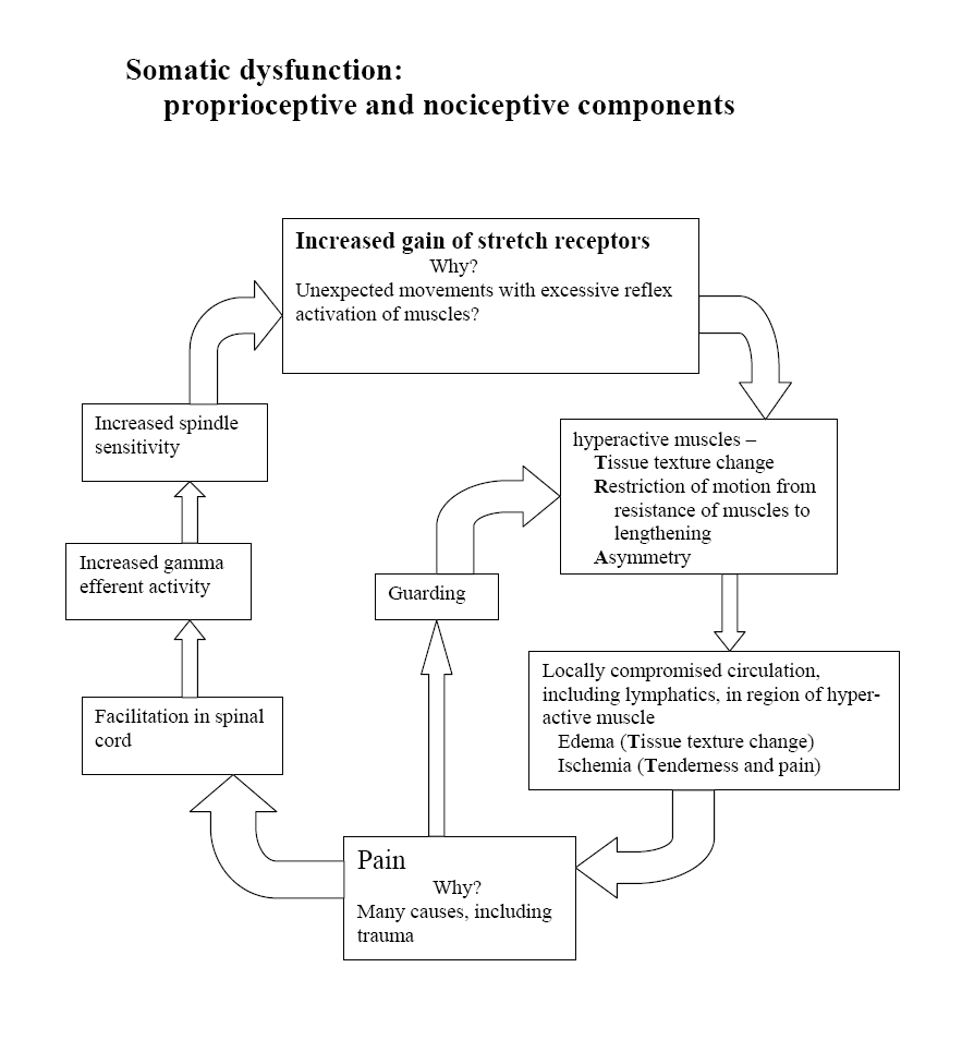
Paragraph Number 17 Somatic dysfunction is defined as “impaired or altered function of related components of the somatic system: skeletal, arthrodial, and myofascial structures and related vascular, lymphatic, and neural elements. (29) Exercise, especially eccentric exercise, commonly results in delayed-onset somatic dysfunction, reduced range of motion, muscle swelling, and tender myofascial points capable of creating local and referred pain. (6) Maximal effort during exercise has also been implicated in the initiation of a variety of overuse phenomena, including somatic dysfunction and myofascial trigger points (MTrPs).

Paragraph Number 18 Osteopathic manipulative treatment (OMT) is most commonly used to reduce or remove somatic dysfunction (29) including MTrPs. OMT and many other hands-on approaches, including massage, physical therapy, chiropractic, and manual medicine treatments, are frequently used in conjunction with more traditional pharmacological approaches to multiple sclerosis. (9) While numerous other reasons involving postulated vascular, autonomic, and nociceptive mechanisms might justify exploring the role of OMT in the treatment of MS subjects, (9) this study is not designed to consider them.

Paragraph Number 19 OMT was paired with MEE in this study for a variety of specific reasons. The palpation of skeletal, arthrodial, and myofascial structures for somatic dysfunction prior to instituting OMT serves to provide secondary data with potential importance should injury or persistent dysfunction arise during the exercise portion of the study. Furthermore, treatment of pain is important in maintaining compliance in exercise studies. The OMT techniques employed in this study were selected to diminish any discomfort associated with somatic dysfunction or MTrPs that might arise from the exercise protocol.

Paragraph Number 20 Regardless of whether an exercise program would introduce somatic dysfunction in this population or not, approximately 55% of the people in this study reported what is called “clinically significant pain” at some time during the course of a lifetime with MS; almost half (48%) were troubled by chronic pain. Another rationale for the MEE/OMT combination to serve as the somatic adjunct in this protocol considers recent studies that suggest a combination back of exercise and manipulation may be superior to exercise alone in back pain. (26)
METHODS


Paragraph Number 21 Seven (7) female subjects between the ages of 42 and 68 years, diagnosed with chronic progressive MS and having an EDSS rating of between 2 and 6, were recruited to participate in this study. All were permitted complete written and oral informed consent according to national standards and those imposed by the Institutional Review Board of the Kirksville College of Osteopathic Medicine. All subjects in this study continued their existing pharmacological care regimen as prescribed by their attending physicians. All subjects participated in an adjunctive specialized somatic care protocol consisting of maximal effort exercise (MEE) program using a three-phase MS IsoPump® exercise protocol in combination with osteopathic manipulative treatment (OMT).

Paragraph Number 22 The IsoPump® is an electrically driven exercise device with which a subject can exert maximum forces through major muscle groups. The proposed effect of these forces is threefold:

1. Increase muscle strength without adding muscle bulk
2. Challenge the muscle component of the arterial system
3. Establish a changed proprioceptive feedback loop by pressure overload

OMT is a form of manual medicine delivered to remove somatic dysfunction and to enhance homeostatic mechanisms.
RESEARCH DESIGN


Paragraph Number 23 The study employed a single blind within subject repeated measures and report design to evaluate the effects of the Isopump® MEE program and OMT over a 12 week period. All seven subjects had the following inclusion and exclusion criteria:

1. were remitted from MS exacerbation for at least six months
2. had been diagnosed with MS for at least two years
3. gave informed consent to participate in this study
4. had no significant spasticity or ataxia
5. had no changes in prescription medicine within previous three months
6. had no clinically diagnosed depression
7. had no pulmonary or bladder infections, or were febrile (>100 deg. F.) at the time of the exercise session.

Paragraph Number 24 The short duration (12 weeks) of the program was intended to minimize maturation while the inclusion and exclusion factors maintain homogeneity of the sample and allowed externalization to the female MS population of a mild (2) to moderate (6) EDSS rating. An independent neurologist conducted the initial neurological evaluation and EDSS rating to insure that the inclusion and exclusion criteria were fully enforced.
MAXIMAL EFFORT EXERCISE INTERVENTION


Paragraph Number 25 The specialized Isopump® exercise program used in this study was a three-phase protocol combining isometric and eccentric vertical leg presses with an isometric semi-erect lunge exercise. In the first two exercise phases the major leg muscles are used while the whole body (leg:torso:arm) is used in the third exercise phase. The exercise protocol was performed twice weekly for twelve weeks. At each session, each individual exercise was performed for 4-6 seconds concomitant with a Valsalva maneuver. Initially, three repetitions were performed in each phase with a minimum rest period of less than 30 seconds between repetitions.

Paragraph Number 26 Frequency of the tests increased as planned from three repetitions of each exercise during the first four weeks, to four repetitions for weeks 5-8, to five repetitions during the last four weeks of the protocol. On the other hand, voluntary duration of exercise effort for all three types of exercise did not change significantly from the start to the end of the exercise intervention period. Exercisers were only capable of exerting maximal forces with Valsalva for an average of 4-8 seconds throughout any of the exercises.

Paragraph Number 27 The Isopump® isometric and eccentric leg exercise phases incorporated a supine anti-orthostatic body position of minus six degrees from horizontal to maximize elevation of torso segment volume. The Isopump® features a visual read-out screen displaying load-cell measurements of the forces applied during all exercises. Strength measured by the load cell was directly recorded every 0.25 seconds by a linked computer for subsequent analysis.
OSTEOPATHIC MANIPULATIVE INTERVENTION


Paragraph Number 28 Twice weekly, after each of the seven subjects exercised, each subject received OMT from an osteopathic physician with special expertise in delivering this somatic intervention. Throughout, the osteopathic physicians involved in the study applied OMT to each subject as determined most individually appropriate. Each OMT session consisted of a variety of techniques as needed to remove somatic dysfunction. This was to maximize axial and appendicular functions, and/or to enhance venous-lymphatic drainage and autonomic functions.
OBJECTIVE MEASUREMENTS


Paragraph Number 29 During interventions, weekly measurements were taken utilizing the Block and Box (BB) test and a Timed 25-foot Walk (TW-25). The Subjective Perception of Fatigue Scale (SPFS) was self-recorded twice weekly before and after each session. Summative evaluations, including the SPFS, TW-25, and BB tests, were conducted at the commencement and completion of the 12-week intervention program as well as at 3, 6 and 9 months after the cessation of the intervention period.

Paragraph Number 30 The SPFS is a seven-item questionnaire assessing features of fatigue on a 7-point Likert Scale; a standard for neurodegenerative disorders. It has demonstrated high test-rated reliability and interval consistency reliability. The BB test counts the number of blocks put into the box in 60 seconds and is a measurement of upper extremity dexterity. The TW-25 simply measures the number of seconds required to walk 25 feet, correlates highly with the EDSS, and also reflects lower extremity strength and endurance.

Paragraph Number 31 Measures of strength (PEAK), endurance (AREA), and duration of maximal effort (TIME) were collected twice weekly by the IsoPump® load-cell during each of the three isometric and eccentric maximal effort exercises. (See Figures 1 for Lunge PEAK. Figure 2 for Isometric Leg Press PEAK. Figure 3 for Eccentric Leg Press AREA). Maximum effort (PEAK) was measured in pounds. Duration of maximal effort (TIME) was measured in seconds. Endurance, indicated by the area under the load-cell generated peak-duration period (AREA), was measured in pound-seconds.
RESULTS


Paragraph Number 32 As reported in the May 2002 JAOA article (30), all individual strength, endurance, ambulation, coordination, and fatigue measures were analyzed to ascertain whether any significant gains were made from Baseline to the end of the 12-week MEE/OMT adjunctive intervention period. Consistent with this study’s additional hypothesis, the statisticians at KCOM and Philadelphia College of Osteopathic Medicine (PCOM) were also asked to evaluate the significance and duration of prolonged effects during a follow-up period without the benefit of further exercise or OMT. The relevant statistics were provided at 3, 6 and 9 months post intervention.

Paragraph Number 33 As previously reported in the (30) univariate analysis by parameter and observation time was conducted calculating arithmetic averages, medians, standard deviations and the associated 95% confidence intervals associated where relevant (see graphs 1-2 and tables 2-3). For each task, a two-factor analysis of variance with repeated measures on both factors test number (see graphs 1-2 and tables 2-3) was used to determine whether there were changes within a session and over the intervention period. Multiple comparisons were performed, when appropriate, using Duncan’s Multiple (DM) Range Test. For the purpose of this study, all participants’ results were assessed from the DM Range Test on five (5) separate occasions: Baseline, Post-Intervention, and at three Follow-Up points timed 3-, 6-, and 9-months after cessation of MEE/OMT.

Paragraph Number 34 Compared to baseline, measures of both strength (PEAK) and endurance (AREA) showed significant improvement (p<0.05) at the end of the 12-week long intervention period as did indicators of quicker ambulation (TW-25) (see graph 1, table 2), improved coordination (BB test) (see graph 2, table 3), and reduction of fatigue (SPFS) as reported in the 2002 JAOA article by Yates, et.al..

Paragraph Number 35 Beyond the original report, this study showed substantial prolonged effects after discontinuing the intervention. Total body strength, as demonstrated by PEAK Lunge (PEAKL) measurements had increased 70% over baseline (p=0.03) and was retained without diminution at all sample points over the nine months following MEE/OMT cessation.

Paragraph Number 36 Although both isometric and eccentric PEAK Leg Press (PEAKLP) measures increased throughout the intervention as previously reported and showed varying degrees of continuing without adjunctive care, the Post-intervention isometric PEAKLP had increased by 36%, Post-intervention eccentric PEAKLP had increased 87%, and, compared to baseline, these eccentric PEAKLP values were significant 3- and 6-months post-intervention. Isometric PEAKLP remained significantly improved for the full 9-month follow-up period.

Paragraph Number 37 As measured by the initial pilot study (30), by the end of the MEE/OMT period, endurance (AREA) increased for both total body lunge (AREAL) and leg presses (AREALP). Endurance measures by the end of the intervention period varied significantly (p<0.05) with isometric AREAL increasing by 93%, isometric AREALP by 113%, and eccentric AREALP by 44%.

Paragraph Number 38 In addition to these substantial strength and endurance improvements, MEE/OMT created a 30.5% reduction (p=0.03) in the Timed 25-foot Walk test. This improvement was fully retained from the end of the intervention throughout the 9-month follow-up period. Dexterity, as measured by the Block & Box test, demonstrated a 13% improvement at six months follow-up for seven subjects.
DISCUSSION


Paragraph Number 39 Overall, research (20,21)suggests that exercise would not be expected to exacerbate MS symptoms except when the physical activity is aerobic and/or performed in hot and humid conditions. (27) Outside these concerns then, MS patients should expect to gain many, if not all of the health-related benefits of an optimally designed exercise protocol. (13)

Paragraph Number 40 Concerning the type of exercise for de-conditioned subjects, a combination of exercise types similar to those selected for this study is recommended over a single isometric or concentric type. Research indicates that a resistance exercise protocol that includes eccentric as well as concentric exercise, particularly when the eccentric exercise is emphasized, results in greater strength gains than concentric exercise alone. (5) Lastayo further demonstrated that significant gains in isometric leg strength were seen in the eccentrically trained subjects only without muscle injury and with minimal increase in metabolic demand for oxygen. 11 (11)

Paragraph Number 41 Brockett demonstrated that continued eccentric exercise of the hamstrings was capable of shifting the optimal angle of human muscle “as a protective strategy” against injury from eccentric exercise. (2) In his study of normal subjects, initial discomfort, swelling, and internal distress caused upon initiation of eccentric exercise disappeared with repeated eccentric training. He and others have postulated that the well-known training effect involves increasing the number of sarcomeres in muscle fibers. (2,14)

Paragraph Number 42 To date most, if not all, studies have failed to conduct substantive follow-up programs to evaluate the prolonged effects of exercise in individuals with MS. There remains a need to conduct follow-up exercise and OMT research to establish what effect each intervention has on the course of Multiple Sclerosis. With the wide variance of symptoms experienced by the subjects, what systemic changes are initiated by these interventions to effect the beneficial changes noted in this pilot study? If MS is an immune dysfunctional response, does exercise and/or OMT provoke the pituitary or endocrine system to facilitate compensatory responses in MS individuals? Further, study is needed to determine whether the whole body (ie. Lunge exercise) is more effective than combined lower extremity exercise (ie. Leg Press). Other questions requiring investigation include whether eccentric exercise has a more lasting effect than isometric type exercise, what interval is most effective between each exercise session, and whether further strength gains are possible with the application of follow-up exercise sessions.

Paragraph 43 The design of the IsoPump® equipment allows the speed of the eccentric exercise to be varied thus altering the resistance applied by the exerciser. This may have important effects for MS sufferers who have bone density loss and who require a longer application of eccentric forces at a slower speed. The current view is that over 60% of MS sufferers may have bone density loss greater than 1.5%. It was observed that the average non-MS exerciser exerts maximal pressure for eight seconds in both isometric and eccentric exercise phases. What fluid and cellular effects does the Valsalva Maneuver maintained for this period of time have on the human mechanism and is this different in individuals with MS?

Paragraph 44 The beneficial effects and strength gains maintained over such a prolonged period of time as found in this study may have applications in a wide range of medical rehabilitation and exercise areas. That strength gains of such a magnitude can be maintained for over six months provoke thought as to the possibility of sustained changes without chemical intervention. Eccentric exercise would appear to be the key to safely and progressively overloading the muscular system and provoking such change. The maintenance of increased strength gains without further exercise sessions offer potential applications for zero gravity situations and increased technique training pre-competition for athletes.


Bibliography

1. Aitkens, S., M. McCory, D. Kilmer, and E. Bernauer. Moderate resistance exercise program: Its effect in slowly progressive disease. Archives of Physical Medicine and Rehabilitation. 74:711-715, 1993.

2. Brockett, C., D. Morgan, and U. Proske. Human hamstring muscles adapt to eccentric exercise by changing optimum length. Med. Sci. Sports Exerc. 33:783-790, 2001.

3. Gehlsen, G., S. Grigsby, and D. M. Winant. Effects of an aquatic fitness program on the muscular strength and endurance of patients with multiple sclerosis. Physical Therapy. 64:653-657, 1984.

4. Goodkin, D., D. Hertsgaard, and J. Seminary. Upper extremity function in multiple sclerosis: improving assessment sensitivity with box-and-block and nine-hole peg tests. Arch Phys Med Rehabil. 69:850-854, 1988.

5. Hilliard-Robertson, P., S. Schneider, S. Bishop, and M. Guilliams. Strength gains following different combined concentric and eccentric exercise regimens. Aviat Space Environ Med. 74:342-347, 2003.

6. Howell, J. Postexercise muscle soreness: a model for the study of somatic dysfunction. Osteopathic Annals. 11:39-45, 1983.

7. Kaufman, M., D. Moyer, and J. Norton. The significant change for the Timed 25-foot Walk in the multiple sclerosis functional composite. Multiple Sclerosis. 6:286-290, 2000.

8. Kraft, G. and A. Alquist. Effect of resistive exercise on strength in patients with multiple sclerosis: Baltimore:Department of Veterans Affairs, Publication 122, 1995, p. 348.

9. Kuchera, M. Osteopathic considerations in neurology. In: Complementary Therapies in Neurology: An Evidence-Base Approach. B. Oken (Ed.) London: Parthenon Publishing, 2004, pp. 49-90.

10. Kurtzke, J. Rating neurological impairment in multiple sclerosis. An expanded disability status scale (EDSS). Neurology. 33:1444-1452, 1983.

11. Lastayo, P., T. Reich, M. Urquhart, H. Hoppeler, and S. Lindstedt. Chronic eccentric exercise: improvements in muscle strength can occur with little demand for oxygen. Am J Physiol Regul Integr Comp Physiol. 276:R611-R615, 1999.

12. Lindsey, J. and J. Wolinsky. IX Demyelinating Diseases. 11 Neurology. ACP Medicine Online. Available at: http://www.acpmedicine.com Accessed, 2005.

13. Medicine, A. C. o. S. Guidelines for Exercise Testing and Prescription. Baltimore, MD: Williams & Wilkins, 1995, 269-287.

14. Morgan, D. New insights into the behavior of muscle during active lengthening. Biophys J. 57: 209-221, 1990.

15. Mosley, P. Exercise, stress and the immune conversation. Exercise and Sports Sciences Reviews. 28, 2000.

16. Petajan, J., E. Gappmaier, A. White, M. Spencer, L. Mino, and R. Hicks. Impact of aerobic training on fitness and quality of life in multiple sclerosis. Ann Neurol. 39:432-441, 1996.

17. Ponichtera, J., T. Mathews, and R. Glaser. Maximal aerobic power of individuals with multiple sclerosis using arm, leg, and combined arm ergometer exercise. Medicine and Science in Sports and Exercise. 24:S73, 1992.

18. Ponichtera, J., M. Rodgers, R. Glaser, T. Mathews, and D. Camaione. Concentric and eccentric isokinetic lower extremity strength inpersons with multiple sclerosis. The Journal of Orthopaedic and Sports Physical Therapy. 16:114-122, 1992.

19. Ponichtera-Mulcare, J. Exercise and multiple sclerosis. Medicine & Science in Sports & Exercise. 25:451-465, 1993.

20. Ponichtera-Mulcare, J. and R. Glaser. Evaluation of muscle performance and cardiopulmonary fitness in patients with multiple sclerosis: Implications for rehabilitation. NeuroRehabilitation. 3:17-29, 1993.

21. Ponichtera-Mulcare, J., R. Glaser, T. Mathews, and D. Camaione. Maximal aerobic exercise in persons with multiple sclerosis. Clinical Kinesiology. Winter: 12-21, 1993.

22. Poser, C. The pathogenesis of multiple sclerosis. Additional considerations. Journal of the Neurological Sciences. 115:S3-S15, 1993.

23. Schapiro, R., J. Petajan, D. Kosich, B. Molk, and J. Feeney. Role of cardiovascular fitness in multiple sclerosis: A pilot study. Journal of Neurological Rehabilitation. 2:43-49, 1988.

24. Somerset, M., R. Campbell, D. Sharp, and T. Peters. What do people with MS want and expect from health-care services? Health Expect. 4:29-37, 2001.

25. Svensson, B., B. Gerdle, and J. Elert. Endurance training in patients with multiple sclerosis. Five case studies. Physical Therapy. 74:1017-1026, 1994.

26. Team, U. B. T. United Kingdom back pain exercise and manipulation (UK BEAM) randomised trial: effectiveness of physical treatments for back pain in primary care. Brit Med J. 329:1381, 2004.

27. Vardy, T. Enhancing homeostasis using osteopathic techniques for multiple sclerosis. Australian Journal of Osteopathy. 8:20-26, 1997.

28. Waksman, B., S. Reingold, and W. Reynolds. Research on Multiple Sclerosis. Desmos. 1:1-2, 1987.

29. Ward, R. C. Foundations for Osteopathic Medicine. 2nd ed. Baltimore: Williams & Wilkins, 2003

30. Yates, H., T. Vardy, M. Kuchera, B. Ripley, and J. Johnson. Effects of osteopathic manipulative treatment and concentric and eccentric maximal-effort exercise on women with multiple sclerosis : A pilot study. J Amer Osteopath Assn. 102:267-275, 2002.
How Osteopathic Manipulative Treatment (OMT) treats Herniated Lumbar Disk, Lumbar Radiculopathy And Mechanical Low Back Pain

Yamamoto New Scalp Acupuncture (YNSA)
Acupoint Frequency In The Treatment
Of Herniated Lumbar Disk, Lumbar Radiculopathy,
And Mechanical Low Back Pain


Richard A. Feely, DO

ABSTRACT


Background Yamamoto New Scalp Acupuncture (YNSA) is an acupuncture microsystem used for treating pain and dysfunction. To date, there has not been a large-scale study of the use, acupoint frequency, or location of YNSA in the treatment of various diagnoses of back pain.

Objective To determine the location frequency of YNSA Basic and Ypsilon points.
Design, Setting, and Patients A retrospective 2-year study of 115 successive patients seen at a private practice who received YNSA: 22 lumbar herniated nucleus propulsus (HNP) cases, 38 lumbar radiculopathy (LR) cases, and 55 somatic dysfunction/low back pain (LBP) cases.

Intervention All patients received osteopathic manipulative treatment (OMT) for identified somatic dysfunction and if pain persisted, YNSA
was used.

Main Outcome Measure Location and frequency of YNSA acupoints to achieve pain relief.

Results A total of 115 patients with back pain had a mean average of 3.17 visits. Ypsilon points most commonly used were Yin of YIN, 86.4%, and the least common were Yang/YANG, 1.6%. The most common Basic points were YIN D1-6, 11.14%, and most common Ypsilon points were left Yin of YIN GB, 3.65%. This treatment approach resulted in mean visits per patient of HNP: 5.27, LR: 2.42, and LBP: 1.82.

Conclusions
The use of YNSA and OMT for low back pain resulted in immediate pain relief with a minimum of needles. YNSA should be further studied for this application.

KEY WORDS

Acupuncture, Yamamoto, YNSA, Scalp, Low Back Pain, Lumbar Radiculopathy, Herniated Lumbar Disk, Ypsilon Points, Osteopathic Manipulation

A heads-up for neck pain sufferers: Try isometrics

A heads-up for neck pain sufferers: Try isometrics
Poor fitness, neglected muscles and bad posture weaken the neck, but a few exercises can help.
By John Briley
Washington Post

December 11, 2006

Today's topic is a pain in the neck. Literally.

Neck pain may seem to stem from a single action — an awkward sit-up, turning your head to see merging traffic or yelling "hi-YA!" while performing martial arts on a mosquito. But for recreational athletes or civilians pursuing a fitter life, these injuries usually stem in part from longer-term neglect.

The culprit, says Dr. Stephen Rice, director of sports medicine at Jersey Shore University Medical Center in Neptune, N.J., is sometimes a poor fitness strategy.

"Many, many people focus [their workouts] on the muscles in the front of their bodies," such as those in the chest, shoulders, abs and biceps, Rice says. But developing those "mirror muscles" while ignoring the muscles that support the spine and torso pulls the body off its preferred balance point on the spine.

Another culprit is bad posture. Many people sit with their shoulders scrunched high, neck craned toward the computer screen and back rounded — for, say, 7 1/2 hours a day. Even a good workout regimen and strong core can do little to neutralize the daily torture.

And so the muscles in and around the neck work harder to keep the head vertical. This continual engagement fatigues the muscles, leaving them vulnerable to strain from even a minor twitch or rotation.

"Your head weighs about the same as a honeydew," Rice says. "If it tilts forward, even 5 degrees, that is a lot of added pressure. Your head won't fall off, but you will use muscle to hold it up."

In proper standing posture, Rice says, "you could drop a plumb line from your earlobe and it would hit your shoulder, hip, knee and ankle." In such alignment, the craftily designed spinal column will support much of the body's weight.

Contrary to what many people are told as children, a ramrod-straight spine is not the goal: The spinal column naturally curves inward at the neck and again in mid-back to help dissipate shock to the vertebrae during impact.

To protect the neck from injury, isometric exercises help build strength.

Do two sets of six to eight reps, twice a week, of the following, placing your hand on your head to provide moderate resistance:

• Lower chin to chest (hand on forehead).

• Raise chin toward ceiling (hand on back of head).

• Ear to each shoulder (hand on side of head).

• Turn head to each side (hand on chin).

Also, slow, light stretching through a normal range of motion helps loosen the neck before a workout. (This is a rare exception to the don't-stretch-a-muscle-that-hasn't-been-warmed-up rule. If anything hurts, stop immediately.)

You'll also want strong core muscles, front and back. Aside from serving as your powerhouse for running, biking and azalea-planting, the core helps support everything above it, including that melon-like noggin.

To self-treat minor strains, rest until it feels better, then try simple stretches (such as the ear-to-shoulder move without resistance). Again: Keep movements slow and painless.

If pain is severe or persists for more than a week, see a doctor. Once healed, regularly stretch the chest and shoulder muscles — to encourage torso balance — and work on that posture: shoulders down and slightly back, head approximately vertical, core firm.

Robert Morris U. advances planned osteopathic program

Robert Morris University's plans for a school of osteopathic medicine have moved ahead with recent approval from the state Department of Education for the school and its doctor of osteopathic medicine degree program.

The university now awaits national accreditation for the school from the American Osteopathic Association's Commission on Osteopathic College Accreditation, and continues to plan to enroll the school's first class in fall 2008.

Working with the accreditation process is the future school's dean, Oliver Hayes, D.O., also vice president of RMU's College of Health Sciences.

The school would be the third osteopathic medical school in the state.

Cervical Mobilization in Post Traumatic Headache/Cervicalgia by By Sherman Gorbis, DO, FAAO

All osteopathic physicians, in their first two years of osteopathic medical college training, are taught Osteopathic Palpatory Diagnosis and Osteopathic Manipulative Treatment (OMT). OMT is one type of manual medicine. OMT is defined as “The therapeutic application of manually guided forces by an osteopathic physician to improve physiologic function and/or support homeostasis that have been altered by somatic dysfunction” (1). Somatic dysfunction is defined as “Impaired or altered function of related components of the somatic (body framework) system: skeletal, arthrodial, and myofascial structures, and related vascular, lymphatic, and neural elements.” Somatic dysfunction is treated using OMT (1). The diagnostic triad for diagnosing somatic dysfunction is ART:

* Asymmetry-determined visually and by palpation
* Restriction of motion-determined by palpation
* Tissue texture abnormality-determined by palpation

Traumatic cervicalgia (neck pain) can occur as a result of a motor vehicle accident (commonly during/after a whiplash injury where the head and neck are thrown forward/backward), sports related injury, or a fall (such as from a ladder, horse, etc).

It would be very common and not un-expected for the patient who has experienced trauma to have Post Traumatic Headache/Cervicalgia (neck pain) with accompanying somatic dysfunction in her/his cervical spine (bones that make up the neck). The cervical spine is divided into two regions based on their motion characteristics:

Atypical cervical vertebrae. This includes C0-C2 (the upper surface of C2). C0 is the occipital bone, which is located in the lower back area of the skull. The occiput articulates (joined together to allow motion between them) with the first cervical vertebra, C1. C1 articulates with the second cervical vertebra, C2. Several muscles that have attachments in the cervical spine, or below, also attach to the occiput. When these muscles become hypertonic (a sustained contraction but not a spasm (as with a ‘charley horse'), headache can occur. If restriction of motion is present between vertebrae, due to a disruption in the motion of the facets (small joints between the vertebrae which have pain generators) neck pain and headache can occur.

The headache may also result from irritation of the right and/or left greater occipital nerve. This travels through the articulation between the occiput and the first cervical vertebra. It then travels up behind the ear and forward along the temple.

Typical cervical vertebrae. This includes C2-T1 (the lower surface of C2-the upper surface of T1). As above, both muscle hypertonicity and motion restriction can lead to headache and neck pain

Once a physician evaluates the patient who has been involved in trauma and he/she has determined that no contraindications (special conditions that render the use of the procedure inadvisable, usually due to risk) exist for manual medicine, the manual medicine provider then has several options. Various types of OMT include:

Soft tissue. This refers is directed toward tissues other than bone or joints (1).

Muscle energy. The patient voluntarily moves the body as specifically directed by the operator; this is from a precisely controlled position against a defined resistance by the operator (1). Muscle energy is, for the most part, directed at loosening tight muscles using isometric (the muscle does not shorten during the contraction) contractions of the affected muscles. It is also directed at providing proper motion of the facet joints between the vertebrae. Facet joints are located both on the inferior and superior surfaces of vertebrae to allow one vertebra to move freely in relation to the vertebra below. Muscle energy techniques can treat facets that are either dysfunctionally ‘opened' or ‘closed'.

Direct Action Thrust (Mobilization with Impulse). Uses a high-velocity/low-amplitude activation, or thrust, to move a joint that is experiencing somatic dysfunction to help restore appropriate physiologic motion (1). This can be, sometimes, accompanied by an audible ‘click' or ‘pop'. However, the goal of treatment is the restoration of motion, not the presence of the sound.

Myofascial Release. This approach engages continual palpatory (the provider's hands in contact with the patient) feedback to achieve release of myofascial (muscles and their soft tissue/fascial coverings) tissues. This can be employed when tissue hypertonicity is present without severe motion restriction.

Other types of OMT include functional indirect and cranio-sacral.

Chiropractic physicians are traditionally trained in the use of Direct Action Thrust. Physical Therapists are well trained in stretching and strengthening exercises; however, more recently, a large number of PT's have been learning the above approaches as well. It would behoove the patient with Post Traumatic Headache/Cervicalgia to have a dialogue with his/her manual medicine provider. The patient should inquire as to the provider's training and the type of modality(s) that might be used and why. Clinical experience has shown that most patients do well when manual medicine is used synergistically with proper stretching/strengthening exercises.

Many insurance companies cover OMT and many osteopathic physicians who include OMT in their practices accept these plans. It is always helpful for the patient to inquire with his/her insurance company, as well as the provider, regarding coverage.
Resources

Ward RC, exec. ed. Foundations for Osteopathic Medicine 2 nd ed. Philadelphia : Lippincott Williams & Wilkins, 2003.
American Academy of Osteopathy (AAO)
This group's mission is to teach, advocate, advance, explore, and research the science and art of osteopathic medicine, emphasizing osteopathic principles, philosophy, palpatory diagnosis and OMT in total health care. Most, if not all, members use OMT in some degree in their practices.
Phone (317) 879-1881
Fax (317) 879-0563
www.academyofosteopathy.org
American Osteopathic Association (AOA)
Patients can inquire about educational materials regarding OMT.
www.aoa-net.org

Sherman Gorbis, DO, FAAO is a graduate of the Kansas City (MO) College of Osteopathic Medicine . He interned at Riverside Osteopathic Hospital ( Trenton , MI ). He earned his certification in Osteopathic Manipulative Medicine (OMM) in 1991 and his Fellowship in the American Academy of Osteopathy (FAAO) in 1995. He is presently an Associate Professor in the Department of OMM at Michigan State University College of Osteopathic Medicine ( East Lansing , MI ).

Muscle Energy Techniques with DVD-ROM (Paperback)

MET is a comprehensive manual therapy system for evaluating and treating joint restrictions of the spine (segmental and intervertebral dysfunctions), rib cage(restricted respiratory motions, dislocations, intraosseous deformities of the ribs), pelvis (sacroiliac, inter-innominate restrictions and dislocations), and extremities (joint restrictions and impairments of muscle length and strength). When you put together all of the elements of somatic dysfunction that MET addresses, there are few manual therapy systems that are as comprehensive and prepared to address structural musculoskeletal dysfunctions and impairments as MET. Possibly because of the name, Muscle Energy has often been misperceived as solely a treatment modality for “tight” muscles. Far too often, MET treatment techniques have been taught without sufficient reference to MET’s distinctive diagnostic algorithms. MET is more than a method of treatment or therapy; it is also a biomechanics-based analytic diagnostic system, using precise physical diagnosis evaluation procedures designed to identify and quantify articular (i.e., joint) range-of-motion restriction. Once we have performed the initial assessment and the articular restrictions have been identified, and the rational sequence for treatment for these restrictions considered, the treatment part of MET can commence. In terms of treatment, “Muscle Energy Technique is a system of manual therapy for the treatment of movement impairments that combines the precision of passive mobilization with the effectiveness, safety, and specificity of reeducation therapies and therapeutic exercise. The therapist localizes and controls the procedures, while the patient provides the corrective forces and energies for the treatment as instructed by the therapist. … MET focuses on joint range-of-motion limitation, and uses light (generally grams or ounces) to moderate force muscular contractions precisely controlled to affect a specific joint, to restore normal joint motion.” (see The Muscle Energy Manual) Since it was originally developed over 45 years ago, MET is now part of the curricula at all of the Osteopathic colleges and physical therapy programs, and is practiced by many Osteopaths, physical therapists, chiropractors, and other manual therapists world-wide.


This comprehensive text describes the basis and practice of Muscle Energy Techniques (MET), a widely recognized approach to treating musculoskeletal dysfunction. It describes those manipulative techniques in which a patient, on request, actively uses his or her muscles from a controlled position in a specific direction against a distinct counterforce applied by the practitioner. These techniques are combined from methods used in physical therapy, osteopathy, chiropractic and manual medicine. A companion DVD-ROM includes video clips demonstrating the application of techniques.

A comparison of osteopathic spinal manipulation with standard care for patients with low back pain.

BACKGROUND: The effect of osteopathic manual therapy (i.e., spinal manipulation) in patients with chronic and subchronic back pain is largely unknown, and its use in such patients is controversial. Nevertheless, manual therapy is a frequently used method of treatment in this group of patients.

METHODS:
We performed a randomized, controlled trial that involved patients who had had back pain for at least three weeks but less than six months. We screened 1193 patients; 178 were found to be eligible and were randomly assigned to treatment groups; 23 of these patients subsequently dropped out of the study. The patients were treated either with one or more standard medical therapies (72 patients) or with osteopathic manual therapy (83 patients). We used a variety of outcome measures, including scores on the Roland-Morris and Oswestry questionnaires, a visual-analogue pain scale, and measurements of range of motion and straight-leg raising, to assess the results of treatment over a 12-week period. RESULTS: Patients in both groups improved during the 12 weeks. There was no statistically significant difference between the two groups in any of the primary outcome measures. The osteopathic-treatment group required significantly less medication (analgesics, antiinflammatory agents, and muscle relaxants) (P< 0.001) and used less physical therapy (0.2 percent vs. 2.6 percent, P<0.05). More than 90 percent of the patients in both groups were satisfied with their care.

CONCLUSIONS:
Osteopathic manual care and standard medical care had similar clinical results in patients with subacute low back pain. However, the use of medication was greater with standard care.

Friday, December 08, 2006

Keeping Kids Healthy: Holistic Medicine - An Alternative Approach

Is acupuncture right for your child? Will it alleviate his asthma? Can hypnosis help cure chronic her frequent headaches? What about his insomnia? Holistic or Integrated Medicine is an approach to medicine seeking to treat the whole person, integrating both traditional and non-traditional forms of medical care. Many doctors are exploring how these non-traditional forms can support traditional methods. Join host Dr. Winnie King as we explore the ways in which non-traditional therapies can work in concert to keep your kids healthy.


Guests:

Roberta Lee, MD - Medical Director, Director of Continuing Education and Co-director of the Fellowship in Integrative Medicine, Center for Health and Healing at Beth Israel Medical Center, New York, NY

Ben Kligler, MD
Associate Medical Director, Center for Health and Healing at Beth Israel Medical Center; Attending Physician, Beth Israel Medical Center; Assistant Professor of family medicine, Albert Einstein College of Medicine, New York, NY; Co-directed the nation's first required residency-training rotation in integrative medicine at Beth Israel Medical Center.

Craniosacral therapy

Spinal tissue rhythm, that is.

Confused?

“People don’t really understand it,” Grinnell readily admits of the new clinic she recently opened at 7451 E. Carson City Road (M-57) just west of Carson City.

The 45-year-old registered nurse has worked at the Carson City Hospital for the past 18 years. After two years of dreaming and planning, she started CranioSacral Therapy, a clinic to treat head, neck and back pain.

Craniosacral therapy is a method of alternative medicine used to assess and enhance the patient’s functioning by accessing the primary respiratory mechanism — the central nervous system’s membranes and spinal fluid.

Grinnell said the practice helps treat chronic headaches, neck and back pain, stress-related discomfort, chronic fatigue, motor coordinator difficulties, eye problems and central nervous system and temporomandibular joint (TMJ) disorders.

She works with Dr. Wesley Lockhart in the emergency room at Carson City Hospital. He inspired her to open the clinic.

“He kind of took me under his wing and he said, ‘I think you’d be good at this,’” Grinnell said. “I’ve always been a hands-on, feel-type person. I listen to the body. I listen to what the rhythm’s telling me and where to start.”

Lockhart works in Carson City on Tuesdays, has a private practice in Lansing and formerly taught at Michigan State University. He uses craniosacral therapy in his practice.

“I think it’ll be a real advantage to this area to have somebody doing the craniosacral therapy,” Lockhart said. “A lot of the people who benefit from it tell me they feel it’s a very deep tissue but indirect treatment. There’s rarely any pain associated with it. It usually just helps relieve things that are deep inside.”

Grinnell’s therapy is all about the right kind of pressure.

“If you put a nickel in your hand, that’s about the amount of pressure I use,” she said.

Grinnell checks for rhythm in a client’s tissue to tell her where the problem lies. Using her hands, she starts with a client’s legs and works her way up to the tailbone and then the head. She holds a client’s head in her hands and listens to the rhythm of spinal fluid to determine whether it’s off-balance.

“I work with the rhythm,” she said. “I overexaggerate the rhythm I’m picking up in your body.

“I just feel like this was something I was meant to do,” Grinnell said. “I really think people need to try it with an open mind and be open to new experiences. It’s very open and if people don’t understand it they can come out and experience it.”

Grinnell is studying with the Upledger Institute in Palm Beach Gardens, Fla., which was founded by John Upledger, an osteopathic physician whose research showed support for the concept of cranial bone movement and cranial rhythm

Difference Between the Letters Behind Doctors' Names

There's a D.O. behind Neil Propst's name, but not all of his patients realize that.

Neil Propst, D.O., says, "Especially, on correspondence that I get, a lot of people just assume that I'm an M.D. That's really common; people just don't think to look."

He says doctors of osteopathy take a more holistic approach to medicine, treating the patient instead of just treating the disease.

A good doctor of any kind will do both, but a D.O. receives extra training in what's called osteopathic manipulative treatment. They perform soft tissue massage and other body manipulations that improve circulation, lymphatic drainage and other systems helping the body heal itself.

Dr. Propst explains, "If a person comes in with a heart attack, I certainly don't think you adjust their back and they get better. I think there is a definite time and place for it. A lot of people come in with chronic back or neck pain - pregnant women especially. There's some manipulations you can do that can kind of help relieve their pain for a time."

Just like M.D.s, D.O.s go to college, medical school and complete a residency program. Then, they both must pass comparable state licensing exams.

As far as medical doctors are concerned, William Blanke, M.D., says, "A D.O., to me, is really not different from a M.D. to be perfectly honest. If anything, I think the D.O. has sometimes a little edge on things - especially in family practice because the D.O.s are more specifically trained. I believe [they] have more experience working the musculoskeletal system."

Unfortunately, Dr. Propst says he doesn't get to do the body manipulations on patients as much as he'd like. Time just doesn't allow it, and that's a problem shared by doctors of every kind.

There are nearly 50,000 D.O.s in the U.S.

A healing touch goes worldwide

A healing touch goes worldwide

RICHARD DYMOND
Herald Staff Writer

LAKEWOOD RANCH - As the holidays approach, five Lake Erie College of Osteopathic Medicine students are thankful for an experience that changed their lives.

The students all traveled to developing countries this year to volunteer their time in hospitals, clinics and refugee camps. And all came away with a gift.

"I really learned how to love and care for someone I never met before," said Jason Wright, 24, a second-year LECOM student from Seattle who traveled to Zambia as part of a Christian Medical Association outreach program.

Wright performed osteopathic manipulation he learned in his first year at LECOM to help a Zambian woman who was experiencing intense neck, lower back and leg pain from carrying a large basket on her head.

"I couldn't always treat them with medicines, but I could place my hands on them," Wright said. "I learned the value of compassionate care."

Second-year student Morganna Freeman lived with a host family in Guatemala for part of this year.

Freeman was taken to a hospital where children infected by AIDS since birth sleep two or three to a bunk.

Freeman recalled reaching into a crib and lifting out a three-year-old girl named Diana, who had shiny black eyes and ringlets of black hair.

"When I picked her up, she put her arms around my neck and didn't let go," Freeman said.

Freeman wasn't prepared for the portion of her medical training she now describes as love.

"Guatemala changed me," said Freeman, who was sponsored by Dartmouth Medical School. "Now I understand why I am doing what I am doing. I want to help those who need me."

The trip has made Freeman want to open clinics herself and mentor future doctors on compassionate care.

"Morgy will be the first doctor in our family," said Freeman's mother, Priscilla, a teacher in San Antonio, Texas, for 28 years. "When she told me she wanted to go to Guatemala I was uncomfortable at first. But now I'm proud of her."

Wright saw patients in shacks and on the street.

He saw 3,000 people in seven days at a copper mine.

"I saw HIV, tuberculosis and sexually transmitted diseases," Wright said. "I saw street kids who huff gasoline to help with their hunger."

LECOM students Aida Kousheshian, 26, Nery Diaz, 42, and Dempsie Morrison, 26, all visited the Dominican Republic this year on a trip sponsored by LECOM's International Medical Society.

"I will always remember a little girl in a hospital who couldn't stop shaking," Morrison said. "She had a fever they were trying to break."

Kousheshian will always remember children swarming around her for stickers of dinosaur airplanes and butterflies, donated by The Toy Lab of Sarasota.

"They put the stickers all over their faces," Kousheshian said.

Diaz, who was born and raised in Cuba, said the Dominican Republic gave her a global vision.

"People we were treating with an anti-parasitic for diarrhea have to go back and drink more contaminated water," she said. "Every developing country needs public health infrastructure."

The students' gifts will make them better doctors, their professors and fellow students said Wednesday.

"Morganna comes into problem-based learning well-prepared every day," said Dr. Emil Adamec, a LECOM professor of neuroscience and pharmacology.

Mark Shank, a fellow student of Freeman's, said Freeman has impressed him with her spirit.

"She has phenomenal leadership skills," Shank said. "She knows when to step up. She's become so confident in herself."

The fact that these students wanted to do something for their fellow man outside of their country is impressive, said fellow student Melissa Roewe.

Clarence "Chip" Colle, associate professor of microbiology and immunology, agreed.

"The choices they made show how compassionate they are about medicine," he said.

Wednesday, December 06, 2006

Mind Body and Spirit: Total Body Healing!

MIND:
Because osteopathy recognizes that all parts of the body work together to create healing, the mind or brain is considered part of this holistic system. Therefore, osteopathy considers that disorder in body structure can cause or exacerbate mental problems like depression. In turn, it is thought that mental disorder can cause or exacerbate physical disease.

BODY:
Osteopathic physicians use all of the tools available through modern medicine including prescription medicine and surgery. They also incorporate osteopathic manipulative treatment (OMT) into their regimen of patient care when appropriate. OMT is a set of manual medicine techniques that may be used to relieve pain, restore range of motion, and enhance the body's capacity to heal.

SPIRIT:
There is no distinct spiritual component of osteopathy, but some modern practitioners recognize a bodily “energy” (like the Eastern concept of chi) as a healing force.

How does osteopathy work?

Long nerves connect the spine to various organs in the body. Andrew Taylor Still believed that every disease or illness began with structural problems in the spine. According to Still, when problems arise in the spine the nerves send abnormal signals to the body's organs. Still called these spinal problems "osteopathic lesions" ("osteo" for bone and "pathic" for diseased), and devised osteopathic manipulation techniques (OMTs) to treat them. Such lesions are detected by the osteopathic doctor from abnormal texture of the skin and other soft tissues of the body as well as from restricted range of motion in the joints. OMTs range from light pressure on the soft tissues to high-velocity thrusts on the joints. These treatments, he believed, would return the nerves to their normal function and allow the blood to flow freely throughout the circulatory system. With structure restored, the body's own natural healing powers would then be able to restore the entire body to full health.

What happens during a visit to the osteopath?

A visit to a D.O. is much like a visit to your family doctor. The D.O. will ask you questions about your medical history, physical condition, and lifestyle. However, because D.O.'s have particular expertise in musculoskeletal systems (namely, bones, joints, and soft tissues like ligaments and tendons), the physical exam of that bodily system will be more extensive than one with your family doctor. During the physical, the D.O. will assess your posture, spine, and balance; check your joints, muscles, tendons, and ligaments; and may use his or her hands to manipulate your back, legs, or arms. Variations in your skin temperature and sweat gland activity will also be measured. If needed, the D.O. will order X-rays and laboratory tests. When the results are in, the D.O. will make a diagnosis and establish a treatment plan for you that may even include prescriptions for medications.

For problems involving the bones, muscles, tendons, tissues, or spine, many current day (but not all) D.O.s use OMTs. There are two categories of OMT procedures: direct and indirect. In direct OMT, "problem" or "tight" tissues are moved (by the D.O., the person being treated, or both) toward the areas of tightness or restricted movement. In indirect OMT, the D.O. pushes the "tight" tissues away from the area of restricted movement, in the opposite direction of the muscle's resistance. He or she holds the tissues in this position until the tight muscle relaxes.

What illnesses and conditions respond well to osteopathy?

OMTs can be applied to a variety of health problems, both musculoskeletal and non-musculoskeletal. According to the US Department of Health and Human Services, OMTs are most effective for back and neck pain. In fact, if you have back pain, you may be able to reduce the amount of pain medication you are taking if you receive OMT as part of your therapy. One study showed that patients with pancreatitis were able to go home from the hospital sooner when they had OMT.

In one small study, people with Parkinson's disease were able to walk better after only one session of OMT. Another study looked at 38 patients who had knee surgery. Those who had OMT were able to walk up stairs 20% earlier than those who did not have OMT.

A study of 100 people with high blood pressure treated only with OMT showed that OMT produced significant reductions in blood pressure.

Studies show that OMT eases breathing, drains the sinuses and relieves the symptoms, duration, and recurrence of the common cold.

Osteopathy may also be an effective way to treat carpal tunnel syndrome. More studies are needed to confirm this.

Examples of other conditions for which OMT may be helpful include:

* stress-related problems (such as tension headaches, muscle spasm)
* strains and sprains (especially of the neck and back)
* shoulder pain
* osteoarthritis
* headaches
* painful menstruation
* injuries (such as whiplash)
* scoliosis (side to side curvature of the spine)
* infantile colic
* insomnia

About Myofascial Release

What is it?
Myofascial release is relatively new. Osteopathic physician Dr. Robert Ward of Michigan State University taught the first course entitled "myofascial release" at that school in the 1970s, and references to it first began to appear in the medical literature in the 1980s. However, as a holistic treatment that looks at the body as an integrated whole, its roots go back a long way, to the soft-tissue manipulations and stretches of osteopathy, which was first done in the nineteenth century.

Myofascial release is a therapeutic treatment utilizing a gentle form of stretching, producing a healing effect upon the body tissues, eliminating pain and restoring motion. Fascia is a connective tissue that surrounds every muscle, bone, nerve, blood vessel, and organ of the body, down to the cellular level. Malfunction of the fascial system due to trauma, posture, or inflammation can create a binding down of the fascia, resulting in abnormal pressure on nerves, muscles, bones, or organs. By freeing up fascia that may be impeding blood vessels or nerves, myofascial release is also said to enhance the body's innate restorative powers by improving circulation and nervous system transmission. People with longstanding back pain, fibromyalgia, recurring headaches, sports injuries, and a host of additional complaints are all said to benefit from the technique

Like many alternative therapies, myofascial release is part of a larger philosophy of healing that emphasizes the importance of mind-body interactions and preventive care. It may also be part of a pain management program that would include behavioral health techniques, acupuncture, drug therapy, nutritional counseling, and relaxation techniques.

How is it practiced?
The therapy's easy stretches break up, or "release," constrictions or snags in the fascia. The stretch is guided by feedback the therapist feels from the patient's body. This feedback tells the therapist how much force to use, the direction of the stretch and how long to stretch. Small areas of muscle are stretched at a time. Sometimes the therapist uses only two fingers to stretch a small part of a muscle. The feedback the therapist feels determines which muscles are stretched and in what order.

Myofascial Pain Syndrome (MPS) and Osteopathic Manipulative Treatment

Myofascial Pain Syndrome (MPS) is a is a painful musculoskeletal condition, a common cause of musculoskeletal pain. MPS is characterized by the development of Myofascial trigger points (TrPs) that are locally tender when active, and refer pain through specific patterns to other areas of the body. A trigger point or sensitive, painful area in the muscle or the junction of the muscle and fascia (hence, myofascial pain) develops due to any number of causes. Trigger points are usually associated with a taut band, a ropey thickening of the muscle tissue. Typically a trigger point, when pressed upon, will cause the pain to be felt elsewhere. This is what is considered "referred pain".


These factors can cause trigger points:

•Sudden trauma to musculoskeletal tissues (muscles, ligaments, tendons, bursae)
•Injury to intervertebral discs
•Generalize fatigue (fibromyalgia is a perpetuating factor of MPS, perhaps chronic fatigue syndrome may produce trigger points as well)
•Repetative motions; Excessive exercise; Muscle strain due to over activity
•Systemic conditions (eg, gall bladder inflammation, heart attack, appendicitis, stomach irritation)
•Lack of activity (eg, a broken arm in a sling)
•Nutritional deficiencies
•Hormonal changes (eg, trigger point development during PMS or menopause)
•Nervous tension or stress
•Chilling of areas of the body (eg, sitting under an air conditioning duct; sleeping in front of an air conditioner)

The fascia is a tough connective tissue which spreads throughout the body in a three dimensional web from head to foot without interruption. The fascia surrounds every muscle, bone, nerve, blood vessel and organ of the body, all the way down to the cellular level. Therefore, malfunction of the fascial system due to trauma, posture, or inflammation can create a binding down of the fascia, resulting in abnormal pressure on nerves, muscles, bones or organs.

This can create pain or malfunction throughout the body, sometimes with bizarre side effects and seemingly unrelated symptoms. It is thought that an extremely high percentage of people suffering with pain and/or lack of motion may be having myofascial problems; but most go undiagnosed, as the importance of fascia is just now being recognized.

Many of the standard tests, such as x-rays, myelograms, CAT scans, eletromyography, etc., do not show the fascia. (John Barnes, P.T., 1992)

Occassionally, trigger points produce autonomic nervous system changes such as flushing of the skin, hypersensitivity of areas of the skin, sweating in areas, or even "goose bumps." The trigger points cause localized pain, although TrPs can involve the whole body.

In three studies, the prevalence of myofascial TrPs among patients complaining of pain anywhere in the body ranged from 30% to 93%; (among patients with chronic craniofacial pain, 55%; and for lumbogluteal pain, 21%.)

The characteristic electrical activity of myofascial TrPs most likely originates at dysfunctional endplates of extrafusal muscle fibers. This dysfunction appears to play a key role in the pathophysiology of TrPs. (Simons 1996)

Subjective shortness of breath can be part of the myofascial pain syndrome of the levator scapulae muscle. In one study, 75 patients who reported neck pain & shortness of breath were examined. Trigger points were located and inactivated with acupuncture needles (dry needling). 68 of the 75 patients in the study reported that their shortness of breath and soreness were abolished immediately after inactivation of the TrPs. The other 7 patients needed a second trial of inactivation. Eliminating the trigger points eliminated the symptoms. (Journal of Muskuloskeletal Pain, 1996)

Like fibromyalgia, Myofascial Pain syndrome is an often misunderstood condition. Even today, some doctors either don't believe that MPS exists or they don't understand its symptoms and treatment.


Treatment of MPS can only begin after an accurate diagnosis is accomplished. Methods for managing this painful condition:

• Trigger Point Therapy {Myofascial release therapy, myotherapy, massotherapy (medical massage therapy)}
• Spray and Stretch technique (stretching of the muscles involved with a vapocoolant spray - a coolant is sprayed on the trigger point to lessen the pain and then the muscle is stretched. this is often done by a physical therapist.)
• Trigger Point Injections (local anesthetic,such as lidocaine, injected directly into the trigger points)
• Dry Needling (the use of a needle without injecting anything)
[TrP injections and dry needling mechanically disrupt the tirgger point. The use of lidocaine is no more effective, but it reduces the soreness afer injection. For MPS there is no role for injected steroids]

• Osteopathic manipulation treatment
• Craniosacral Therapy

• Physical Therapy (hands-on)
• Exercise
• Improvement of nutrition
• Changing sleeping habits
• The use of tricyclic antidepressants in low doses
• Elimination of stress; Biofeedback; Counseling for depression that may result from this painful condition

An active trigger point when treated well or with rest will become latent (quiet, or not causing active symptoms). It can often resurface after trauma after acute overload or fatigue, or even sudden exposure to cold. Conversely, new trigger points may arise elsewhere, or at least become more sinificant as others become latent.

For MPS, you should see a doctor knowledgeable in chronic pain such as a osteopathic physical medicine doctor (a physiatrist), or a osteopathic neurologist. The diagnosis is made by the history and physical exam. There is no lab test nor imaging studies to confirm the diagnosis. A history of acute trauma or chronic overuse should be looked for.. On exam, there is typically restricted motion with pain of the affected muscle. Other medical problems need to be ruled out with imaging or other studies. For instance, if a patient presents with back pain, disc and other problems need to be ruled out.

Altered Pain Perception Accompanies MPS: A Danish study indicates that people with chronic myofascial pain perceive and transmit pain differently than people without the syndrome. As many as 72 percent of people with fibromyalgia may have trigger points associated with myofascial pain.

Source: "Qualitatively altered nociception in chronic myofascial pain," by L. Bendtsen, R. Jensen, and J. Olesen, Pain, 65 (1996), pages 259-264